
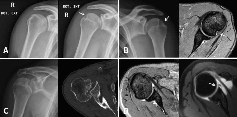
Figure 5. A and B: two cases of patients with Hill-Sachs lesions. A: comparison showing that it is easier to visualize the defect with the shoulder in internal rotation, especially in the case of a small defect; B: the same Hill Sachs lesion is observed on a comparative basis in the plain radiograph and MRI scan; C: computed axial tomography (CAT) view of a reverse Hill-Sachs lesion; D: ALPSA type lesion in conventional magnetic resonance imaging (MRI) and arthro-MRI. The latter technique is useful in the event of diagnostic doubt, since it offers more precise information about the type and characteristics of the capsulolabral lesion.