
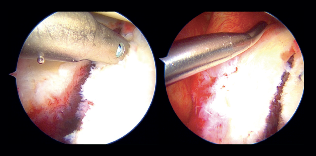
Figure 3. Arthroscopic view of a right shoulder. Position in left lateral decubitus. Anterosuperior vision portal. The image at left shows placement of the most distal implant in the classical Porcellini-Sugaya technique. The image at right shows passage of the suture through the anteroinferior labrum.