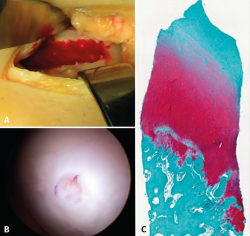
Figure 4. A: extensive chondral lesion of the internal condyle covered by the clot after augmented microfractures with chitosan based hydrogel; B: arthroscopic appearance of the totally covered zone and biopsy sampling orifice after 13 months; C: histological appearance of the biopsied zone. Note the "hyaline" appearance of the tissue and good interdigitation with the subchondral bone. Image of the randomized study with BST-CarGel.
