
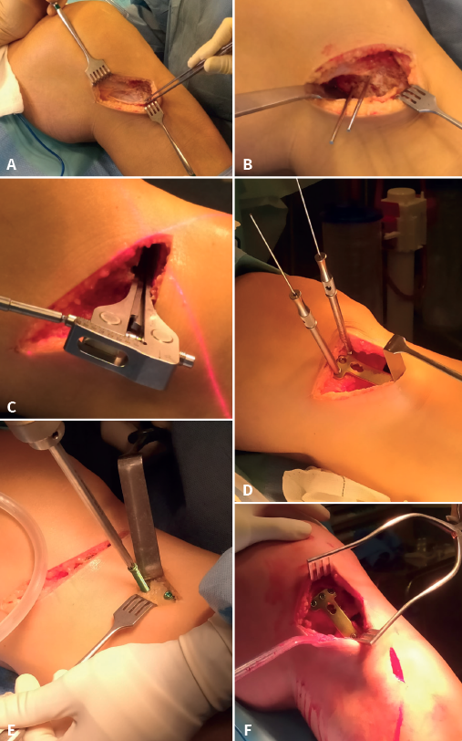
Figure 2. Medial osteotomy surgery. A: medial approach, direct visualization of the medial collateral ligament (MCL) and goosefoot; B: needles placed as guides for the horizontal arm of the osteotomy; C: calibrator of the osteotomy angle after opening with the chisels; D: plate fixation of the opening, after wedge positioning; E: placement of distal screws; F: final plate position.