
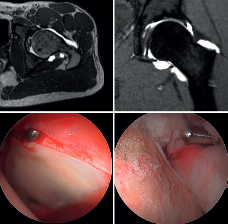
Figure 2. Combined femoroacetabular impingement in a middle-aged male patient. At top, axial and sagittal views of the preoperative MRI study, showing a deformity at the head-neck transition (left) and a chondrolabral junction lesion with excess coverage in the upper area (right). At bottom, intraoperative details of the chondrolabral junction lesion (left) and correction of the femoral deformity (right).