
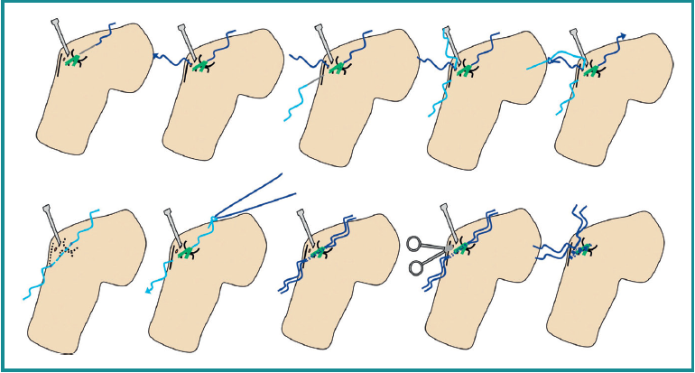
Figure 9. Schematic drawing of the technique used. From the top left corner to the right. An Abbocath is entered in the lateral epicondyle. It is directed intra-articular to the soft spot and a PDS suture is advanced through it into the joint. The distal end is retrieved at the soft spot. A second suture is inserted into the joint in the same way from the subcutaneous border of the ulna, at the level of the origin of the lateral collateral ligament at the supinator crest. It is retrieved through the soft spot and both sutures are knotted. Traction is applied to one end to leave a single suture running from the lateral epicondyle, intra-articular deep to the lateral capsule to the origin of the lateral complex at the supinator crest and the subcutaneous border of the ulna.
A second suture is folded in half, and the intra-articular suture is used as transport suture. Mosquito forceps are used to retrieve the ends of the sutures at the level of the soft spot for knotting. Illustration courtesy of Dr. van Riet and the MoRe Foundation.