
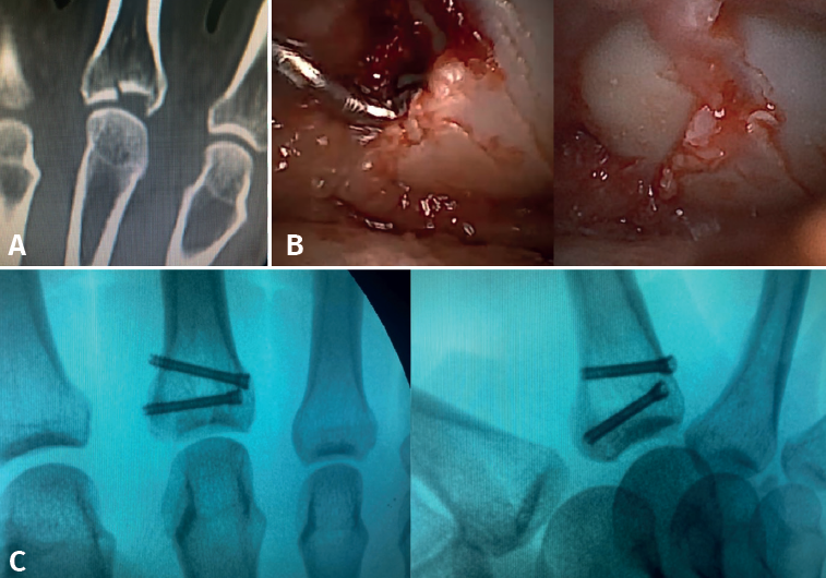
Figure 8. Fracture of the base of P1 of a third finger. Axial compression mechanism causing collapse of a central fragment. A: computed tomography images showing a large collapsed central fragment; B: arthroscopic visualization allows us to perform adequate reduction with the aid of a palpator and Kirschner wires through an extra-articular bony window; C: the size of the fragments and the characteristics of the fracture allow us to perform correct osteosynthesis with cannulated screws, which also accelerates the recovery process.