
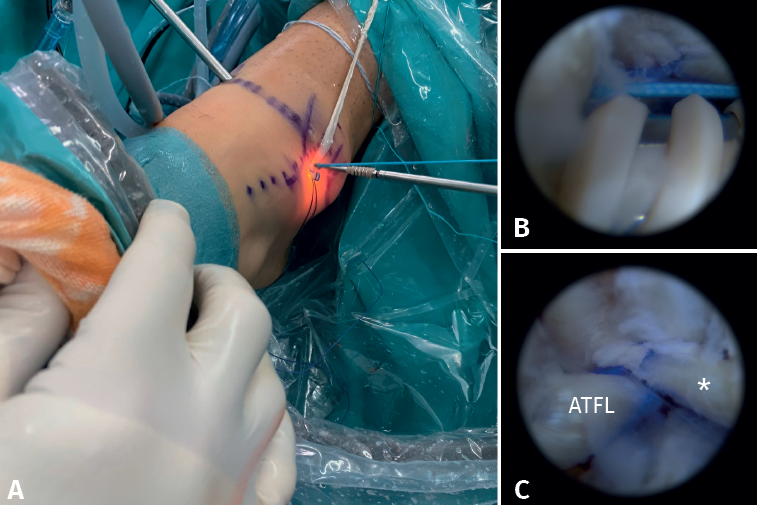
Figure 10. A: external view of insertion of the implant at talar level through the accessory anterolateral portal; B: arthroscopic view of insertion of the implant in the blind talar tunnel; C: arthroscopic view of the anterior talofibular ligament (ATFL) and the fascicle that we will use for the calcaneofibular ligament (CFL) (*).