
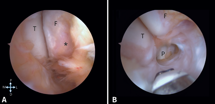
Figure 3. Arthroscopic camera view through the medial portal. A: the lateral groove between the talus (T) and the fibula (F) is observed. We can see the denuded footprint of the ATFL (*); B: the tibia and fibula can be seen, and the peroneus longus (P) is marked at the bottom of the image.