
Clinical-functional and radiographic outcomes of medial valgus-producing tibial osteotomy
Resultados clínico-funcionales y radiográficos de la osteotomía tibial de apertura medial valguizante
Introduction
Gonarthrosis is a chronic degenerative condition of the knee(1). It is increasingly common and poses an important health problem for the current population(2), particularly due to the increase in life expectancy and obesity(3). The degree of osteoarthrosis is defined both clinically and radiologically(1). The medial compartment is most often affected, causing varus gonarthrosis. In order to prevent the progression of this kind of deformity, use can be made of medial valgus-producing tibial osteotomy, which realigns the knee and redistributes loading(2). It has been shown that this procedure alleviates pain and improves the function of the knee, particularly in middle-aged patients, affording good quality of life and delaying and/or avoiding the need for knee replacement surgery(4). In relation to other types of osteotomies performed as treatment for gonarthrosis (e.g., external subtraction osteotomy), valgus-producing tibial osteotomy avoids possible damage to the external popliteal sciatic nerve, keeps the proximal tibiofibular joint intact, preserves bone stock, and is technically more simple to perform(5).
The present study reviews the clinical-functional and radiological outcomes of medial valgus-producing tibial osteotomy in a sample of patients, and explores whether these outcomes have an impact upon patient quality of life and pain perception, using as parameters the body mass index (BMI), the time elapsed from surgery, and the degree of correction of the mechanical axis.
Material and methods
A retrospective observational study was carried out in patients subjected to valgus-producing tibial osteotomy due to knee arthrosis caused by genu varum, between April 2014 and December 2019.
A total of 35 osteotomies in 31 patients (5 females and 26 males) were carried out. The mean patient age was 50.5 years (range 40-63). Patient follow-up extended from the date of surgery to December 2020, with a mean duration of follow-up of 43.7 months (range 12-80). The study sample consisted of consecutive patients.
The sample characteristics are reported in Table 1.
reacae.30380.fs2201001en-table1.png

Table 1. Sample characteristics.
The inclusion criteria were: patients with an active lifestyle, varus gonarthrosis, Ahlbäck II-III, with pain in the compartment, limiting quality of life, no involvement of the external compartment, the absence of associated comorbidities (such as inflammatory joint disorders) and BMI < 40 kg/m2.
Quality of life after surgery was assessed based on the Knee Injury and Osteoarthritis Outcome Score (KOOS) for each operated knee(6).
Preoperative planning
The method of Miniaci was used to plan osteotomy(7,8,9). In the teleradiographic study of the lower extremities, we traced the real mechanical axis (or Mikulicz line, extending from the center of the femoral head to the center of the ankle) and the desired mechanical axis (in our case we used the Fujisawa point, at 62.5% of the total width of the tibial plateau, measured from its medial margin)(10). Taking as vertex a point located 1 cm below the external joint line, immediately above the head of the fibula, we traced two lines to the ankle, joining with the two aforementioned axes traced at that level. This yielded the alpha correction angle (α), which represents the opening wedge to be used to obtain the desired postoperative correction (Figure 1).
reacae.30380.fs2201001en-figure1.png

Figure 1. Preoperative planning with teleradiography. A: blue line: real mechanical axis or Mikulicz line. Green line: desired mechanical axis. Green dot: Fujisawa point; B: using these two lines we obtain the alpha correction angle (α), which yields the wedge angle to be used in the osteotomy.
Surgical technique
All the patients were operated upon by the same surgeon, with use of the standard TomoFix® plate as osteosynthesis material.
reacae.30380.fs2201001en-figure2.png

Figure 2. Medial osteotomy surgery. A: medial approach, direct visualization of the medial collateral ligament (MCL) and goosefoot; B: needles placed as guides for the horizontal arm of the osteotomy; C: calibrator of the osteotomy angle after opening with the chisels; D: plate fixation of the opening, after wedge positioning; E: placement of distal screws; F: final plate position.
Osteotomy was performed adopting a medial oblique approach. The pes anserinus (goosefoot) and anterior most portion of the inner lateral ligament (ILL) were partially deinserted in order to secure wide exposure of the proximal tibial epiphysis. Then, two parallel 3-mm needles (one anterior and the other posterior) were inserted from 3-4 cm below the medial joint line to the lateral point used as vertex of the correction angle. These two needles served as guides for the horizontal arm of the osteotomy. In the anterior portion, at the level of the anterior tibial tuberosity (ATT), we performed the osteotomy perpendicular to the anterior needle, posterior to ATT and guided proximally. After completing the cutting sequence, the chisels were inserted without reaching the external cortical layer, followed by the distractor, until achieving opening of the osteotomy with the planned α angle. Following the above, the graft wedge was inserted in the created opening ChronOS®) and was fixed with a TomoFix® plate screwed proximal and distal (both from DePuy Synthes) (Figures 2 and 3).
reacae.30380.fs2201001en-figure3.png

Figure 3. Immediate postoperative radiological control.
The postoperative protocol included early mobilization, avoidance of weight bearing for 6 weeks, and periodic teleradiography controls to check consolidation.
Statistical analysis
A Microsoft Excel® (version 18) database was created for comparing the results of the KOOS and the time from surgery, postoperative BMI, and degree of correction of the axis. The IBM SPSS version 28.0 statistical package was used to analyze the data. The values were reported as the mean ± standard deviation (SD). Data normality tests were performed Parametric tests (Pearson correlation coefficient) were used in the case of variables showing a normal distribution, while nonparametric testing (Spearman correlation coefficient, ρ) was used for variables with a non-normal distribution.
In the statistical program, the values of the coronal axis were expressed as negative values for varus and positive values for valgus.
Results
Of the 31 patients (35 knees) initially included in the study, only 28 (32 knees) were evaluated at least one year after surgery (91% follow-up rate).
The results were classified into 5 subscales (symptoms, pain, activities of daily living, sports activities and quality of life) within the KOOS. Specifically, the symptoms score was 88.5, the score for pain was 76, activities of daily living 74.3, sports activities 53.8, and quality of life 67.5 (Table 2).
reacae.30380.fs2201001en-table2.png

Table 2. KOOS outcomes.
Only two patients required total knee replacement surgery (TKR) due to the persistence of pain in the internal compartment after the initial osteotomy: one patient at one year postsurgery, and the other after two years.
Table 3 reports the mean values corresponding to the radiological findings of the sample: correction of the femorotibial angle or anatomical axis (postoperative valgus values of 5-7º being regarded as normal), reduction of the distance of the mechanical axis after the operation, and the Insall-Salvati index for the patella. Among the 35 osteotomies, the presurgical mean anatomical femorotibial angle was 2.5º varus, and the postsurgical mean axis angle was 4.13º valgus. Of the 34 patients, 53% (n = 18) achieved a valgus femorotibial angle of between 5-7º. However, although only approximately one-half of the patients reached the valgus target of between 5-7º, the mean difference between the pre- and postoperative axes was 6.6º (p < 0.001).
reacae.30380.fs2201001en-table3.png

Table 3. Analyzed pre- and postoperative data.
We also measured the distance of the mechanical axis of each knee to the Fujisawa point, before and after surgery. This distance was seen to decrease secondary to varus reduction of the operated knees. Specifically, from the initial mean distance of 34.13 mm, a mean reduction to 14 mm was achieved (with a mean reduction of 22.22 mm).
The mean pre- and postoperative Insall-Salvati index values were 0.88 and 0.84, respectively. The mean of the differences between the pre- and postoperative index values was 0.03. This difference was statistically significant.
After analyzing the clinical and radiological outcomes, we explored whether there was a relationship between the KOOS scores and the time elapsed from surgery, postoperative BMI, and the degree of correction of the mechanical axis after surgery.
- With regard to the follow-up time from surgery, the results of the KOOS were correlated for each subcategory to the follow-up time of the 32 osteotomies. No statistically significant correlation (p<0.05) was found between the time from surgery and the results of the KOOS questionnaire. This was a negative correlation, i.e., the longer the time from surgery, the lower the KOOS score.
- In relation to BMI, no correlation was found between this index and the results of the KOOS questionnaire.
- In the case of the postsurgical valgus axis, we analyzed the correlation between the clinical results (mean total KOOS score of the patients) and the degree of postsurgical correction achieved. This was a positive correlation, i.e., the greater the postsurgical valgus axis (greater degree of total correction), the greater the KOOS score and patient satisfaction. Although a clinical correlation was observed, statistical significance was not reached (p>0.05).
- We also examined whether the result varied partially in some of the individual items, and in this regard the mean KOOS score of the subcategory corresponding to activities of daily living proved significant.
Discussion
The present study was carried out to determine whether varus of the knee is related to an improved clinical outcome of the patient and whether this association worsens over time.
From the biomechanical perspective, it is to be expected that a biplanar osteotomy such as that described in this study will result in a decrease in patellar height secondary to distalization of the anterior tibial tuberosity (ATT)(11), since an augmentation wedge is involved, and the cutting design produces a slight descent of the patella - a situation that theoretically could have a negative impact upon the functional outcomes of the surgery. According to the literature(12), this is not a factor considered in all studies, since in general the height of the patella is not measured before osteotomy, and likewise no evaluation is made of whether this decrease in height has functional repercussions for the patient(13).
In our study we measured the height of the patella, with a pre- and postoperative Insall-Salvati index of 0.88 and 0.84, respectively. Our osteotomy had no functional impact upon the height of the patella (the mean difference between the pre- and postoperative index was 0.03). However, it did exert a clinical effect, since the lowest KOOS score corresponded to the sports activities subscale (reaching statistical significance), with items exploring squatting or kneeling function. Thus, our patients, in coincidence with the data found in the literature(14,15), presented limitation of activities that increase stress upon the femoropatellar joint.
In their study, Westrich et al. found that 80% of the patients subjected to total knee replacement surgery and previously subjected to valgus-producing osteotomy presented a low patella. This increases the surgical time and the difficulty of total knee replacement, and moreover produces greater postoperative femoropatellar pain(16). LaPrade et al.(17) confirmed that arthrotomy for performing total knee replacement can become complicated following a tibial osteotomy with distalization of the patella. This did not occur in our two patients that required reconversion to total knee replacement surgery.
In our opinion, it seems logical that in those patients with a clearly low patella, consideration should be made of an alternative technique such as lateral subtraction osteotomy, which produces the opposite effect, i.e., ascent of the patella. In order to avoid this complication, the literature mentions a number of techniques, such as ATT distalizing osteotomy, which has been shown to reduce femoropatellar impingement and maintains alignment(18).
With regard to the clinical results obtained, the patients experienced improvement particularly in relation to pain and the clinical symptoms, as also reported in the literature(19,20). The cohort study published by Bastard (21) confirmed the hypothesis that valgus-producing tibial osteotomy allows the patients to return to normal life after one year, with improvement of the pain and quality of life as measured with the SF-36 questionnaire. In our study, a statistically significant association was observed between the KOOS score referred to activities of daily living and the degree of varus correction, i.e., our patients did not experience discomfort or complications in their daily life that prevented them from continuing their routine activities.
Surgical success above all depends on adequate patient selection(20). The ideal patient for medial osteotomy is an active individual (but not involved in activities like jumping or running) of young age (40-60 years), with isolated pain of the medial joint line, a BMI of < 30 kg/m2, full mobility of the knee, osteoarthrosis in the internal compartment, with Ahlbäck I-II, and almost normal femoropatellar and lateral compartments(22,23,24).
With regard to the correction objective, there is general agreement that mild valgus overcorrection affords better outcomes in osteotomy. However, the specific degree of valgus correction remains to be established(20,25).
In our sample, although only approximately one-half of the patients reached the valgus target of between 5-7º, the mean difference between the pre- and postoperative axes was 6.6º (p < 0.001)(Table 4), which means that our patients achieved an average valgus improvement of 6.6º. In other words, statistically significant correction was achieved, demonstrating that the surgery exerts a valgus-producing effect upon the axis, and thus reduces pressure on the internal compartment.
reacae.30380.fs2201001en-table4.png

Table 4. Femorotibial angle and distances to the mechanical axis.
Hee-June Kim(26), reported a correction of the tibiofemoral angle from varus 8.8º to valgus 2.1º after surgery (p < 0.001). Similar correction outcomes were obtained by Da Silva et al.(27) (from preoperative varus 11.6º to postoperative valgus 4.3º) and Morin et al.(28) (preoperative varus 7º to valgus 3º), with good clinical results. Nevertheless, the literature on the biomechanics following valgus-producing osteotomy remains insufficient. The relationship between the degree of joint degeneration and the desired alignment remains the subject of debate.
The TomoFix® used is a locking compression plate (LCP) that affords rigid fixation and early rehabilitation in the postoperative period(29,30,31), with greater stability than other osteotomy fixation devices(32). However, the plate had to be removed in 5 of our patients due to intolerance issues.
As limitations of the present study, mention must be made of the modest sample size and the lack of certain data such as the preoperative KOOS scores. On the other hand, 6 of the patients had a follow-up period of less than two years. A longer follow-up of patients of this kind would be needed in order to assess the long term outcomes of this surgery, the changes in quality of life, or the total knee replacement conversion rate.
Conclusions
The present study evaluated the clinical-functional and radiological outcomes of medial valgus-producing tibial osteotomy, evidencing subjective improvement of the patient basal condition, together with valgization of the mechanical axis. The patients showed good outcomes over short to middle term follow-up.
Tablas
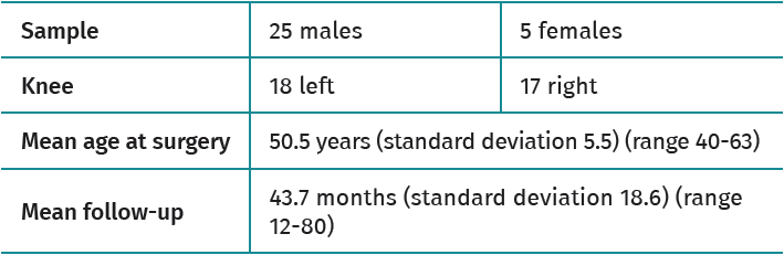
Table 1. Sample characteristics.
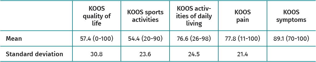
Table 2. KOOS outcomes.
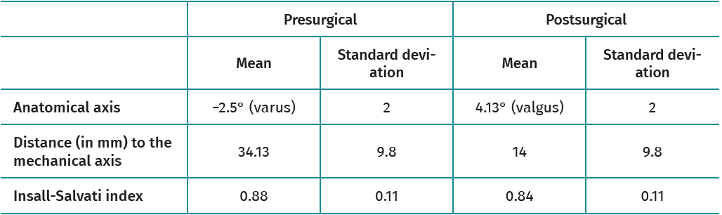
Table 3. Analyzed pre- and postoperative data.
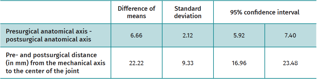
Table 4. Femorotibial angle and distances to the mechanical axis.
Figuras
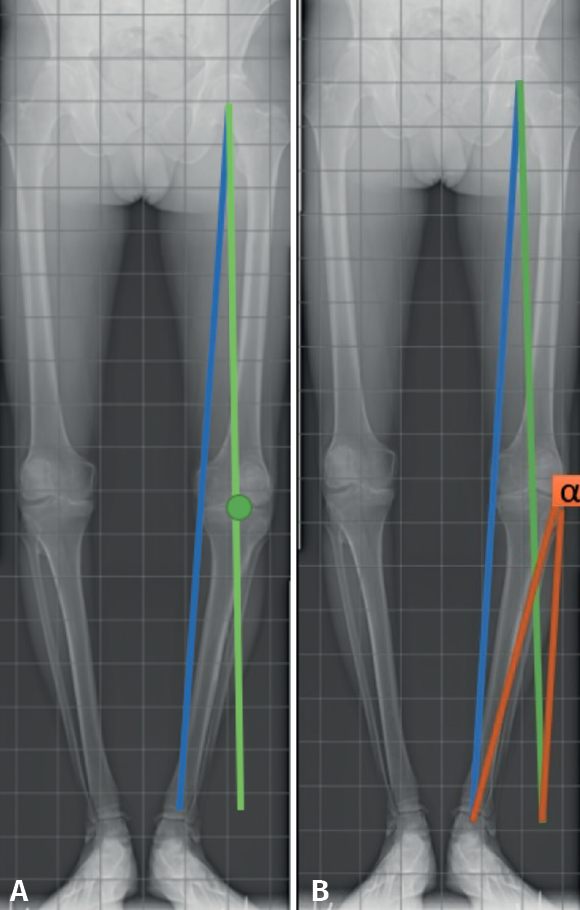
Figure 1. Preoperative planning with teleradiography. A: blue line: real mechanical axis or Mikulicz line. Green line: desired mechanical axis. Green dot: Fujisawa point; B: using these two lines we obtain the alpha correction angle (α), which yields the wedge angle to be used in the osteotomy.
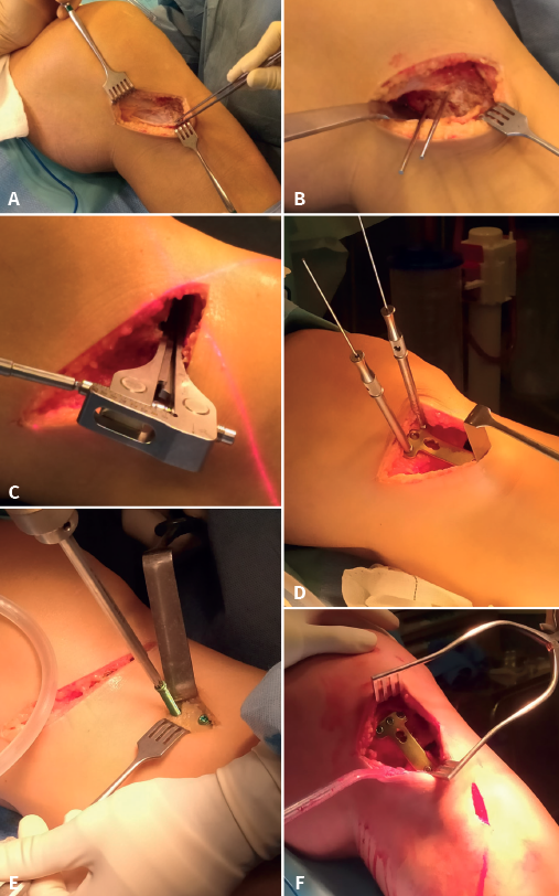
Figure 2. Medial osteotomy surgery. A: medial approach, direct visualization of the medial collateral ligament (MCL) and goosefoot; B: needles placed as guides for the horizontal arm of the osteotomy; C: calibrator of the osteotomy angle after opening with the chisels; D: plate fixation of the opening, after wedge positioning; E: placement of distal screws; F: final plate position.
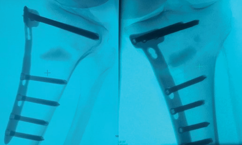
Figure 3. Immediate postoperative radiological control.
Información del artículo
Cita bibliográfica
Ethical responsibilities
Conflicts of interest. The authors state that they have no conflicts of interest.
Financial support. This study has received no financial support.
Protection of people and animals. The authors declare that this research has not involved human or animal experimentation.
Data confidentiality. The authors declare that the protocols of their work centre referred to the publication of patient information have been followed.
Right to privacy and informed consent. The authors declare that no patient data appear in this article.
Descargar artículo:
Licencia:
Este contenido es de acceso abierto (Open-Access) y se ha distribuido bajo los términos de la licencia Creative Commons CC BY-NC-ND (Reconocimiento-NoComercial-SinObraDerivada 4.0 Internacional) que permite usar, distribuir y reproducir en cualquier medio siempre que se citen a los autores y no se utilice para fines comerciales ni para hacer obras derivadas.
Comparte este contenido
En esta edición
- From the start to the top: the history of knee osteotomies
- Clinical-functional and radiographic outcomes of medial valgus-producing tibial osteotomy
- Patellar tendinopathy: a practical consensus guide for choosing the best treatment
- Evolution of spinal endoscopy. Where do we come from and where are we going?
- Partial ruptures of the rotator cuff
- Arthroscopic core decompression of the femoral head with autologous stem cell placement as treatment for femoral head osteonecrosis
- Extraarticular plasty as sole maneuver in the correction of rotational instability due to rupture of the anterior cruciate ligament in an elderly patient
- Fully arthroscopic osteosynthesis with button placement in acute anterior glenoid fracture: a case report
- Arthroscopic visualization of the popliteal hiatus and localization of an intraarticular free body
Más en PUBMED
Más en Google Scholar
Más en ORCID

Revista Española de Artroscopia y Cirugía Articular está distribuida bajo una licencia de Creative Commons Reconocimiento-NoComercial-SinObraDerivada 4.0 Internacional.